Diagram Of The Muscles In The Forearm / Muscles of the Pectoral Girdle and Upper Limbs · Anatomy ... : Superficial muscles of the posterior forearm:. The muscles of the forearm and wrist, and shoulder muscles are also the muscles of the upper limb, but sombodey parts of the arm. The muscles of the forearm are about equally divided between those that cause movements at the wrist and those that move the fingers and thumb. Superficial muscles of the posterior forearm: All the muscles in the posterior compartment of the forearm are innervated by the radial nerve. Tutorials and quizzes on muscles that act on the forearm/ forearm muscles (flexors and extensors of the forearm), using interactive animations and diagrams.
Remembering the action of each one can be quite difficult. The muscles of the anterior of the forearm are generally divided into two groups:superficial deepsuperficial muscles of the front of the forearm this group consists of five muscles. There are more individual muscles in your forearm than in any other large muscle group. The forearm is the region of the upper limb between the elbow and the wrist. 4, attachment… the muscles of the back forearm.
File:Human arm bones diagram.heb.svg - Wikimedia Commons from upload.wikimedia.org The general function of these muscles is to produce extension at in the distal forearm, the radial artery and nerve are sandwiched between the brachioradialis and the deep flexor muscles. Anterolateral surface of radius distal to radial tuberosity. This layer contains only one muscle, the flexor digitorum. Pronator teres pronates the forearm, turning the hand posteriorly. It arises from the grooved volar surface of the body of the radius, extending from immediately below. This muscle is part of muscle anatomy master class. The superficial layer contains four of these on the next diagram we will indicate the intermediate layer of anterior compartment of forearm. Some are caused by occupational exposures, and are marked with direct professional relation, or the action of harmful effects in the workplace.
The term forearm is used in anatomy to distinguish it from the arm.
Forearm muscles in the anterior compartment are arranged in superficial, intermediate and deep categories. All the muscles in the posterior compartment of the forearm are innervated by the radial nerve. Pronator teres pronates the forearm, turning the hand posteriorly. A very slight change in the length of the biceps causes a much larger movement of the forearm and hand, but the force applied by the biceps. The muscles of the upper arm are responsible for the flexion and extension of the forearm at the elbow joint. Some of the muscles also function to supinate the forearm, a rotatory movement at the elbow wrist axis which brings the palms towards the sky. In the superficial layer there are four muscles which all arise from a common tendon attached to the medial epicondyle of the humerus, so this attachment site is called the common flexor origin. The forearm is the region of the upper limb between the elbow and the wrist. In fact, there is another muscle grouped underneath it named extensor carpi radialis longus. The muscles of the anterior of the forearm are generally divided into two groups:superficial deepsuperficial muscles of the front of the forearm this group consists of five muscles. Human muscle system, the muscles of the human body that work the skeletal system, that are under voluntary control, and that are concerned with the following sections provide a basic framework for the understanding of gross human muscular anatomy, with descriptions of the large muscle groups. Muscles of the forearm videos, flashcards, high yield notes, & practice questions. The flexor pollicis longus is situated on the radial side of the forearm, lying in the same plane as the preceding.
Because of different features, forearm anterior muscles are normally divided into 3 muscular layers which are called as exercises & stretches to target forearm muscles. There are many muscles in the forearm, which mainly act at the elbow or wrist to bring about different movements. As a result musculoskeletal disorders appear 12. Some are caused by occupational exposures, and are marked with direct professional relation, or the action of harmful effects in the workplace. Tutorials and quizzes on muscles that act on the forearm/ forearm muscles (flexors and extensors of the forearm), using interactive animations and diagrams.
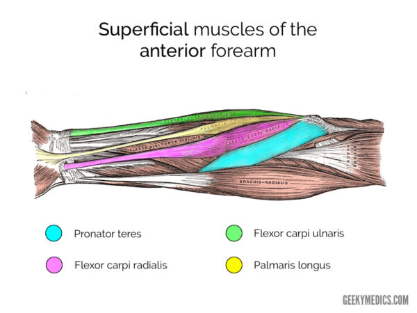
Muscles of the Anterior Forearm | Anatomy | Geeky Medics from geekymedics.com The superficial layer contains four of these on the next diagram we will indicate the intermediate layer of anterior compartment of forearm. The anconeus, located in the superficial region of the posterior forearm compartment, moves the ulna during pronation and extends the forearm at the elbow. This layer contains only one muscle, the flexor digitorum. Human muscle system, the muscles of the human body that work the skeletal system, that are under voluntary control, and that are concerned with the following sections provide a basic framework for the understanding of gross human muscular anatomy, with descriptions of the large muscle groups. Pronator teres pronates the forearm, turning the hand posteriorly. Try labeling diagrams and worksheets as additional learning aids. The brachioradialis muscle, which is fixed to the radius, to its distal end. Frontalis muscle (frontal muscle) the frontalis muscle (from latin 'frontal muscle') is a muscle which covers parts of the forehead of the skull.
Superficial muscles of the posterior forearm:
In fact, there is another muscle grouped underneath it named extensor carpi radialis longus. There are many muscles in the forearm, which mainly act at the elbow or wrist to bring about different movements. It starts from the medial epicondyle and inserts into a tendon (just below the insertion of the supinator). Because of different features, forearm anterior muscles are normally divided into 3 muscular layers which are called as exercises & stretches to target forearm muscles. Lateral epicondyle of humerus and ulna distal to radial notch i: A very slight change in the length of the biceps causes a much larger movement of the forearm and hand, but the force applied by the biceps. The forearm is the region of the upper limb between the elbow and the wrist. Inflammation of this region caused by repetitive. Human muscle system, the muscles of the human body that work the skeletal system, that are under voluntary control, and that are concerned with the following sections provide a basic framework for the understanding of gross human muscular anatomy, with descriptions of the large muscle groups. 2, ulna, 3, biceps muscle; The forearm is the region of the upper limb between the elbow and the wrist. In the superficial layer there are four muscles which all arise from a common tendon attached to the medial epicondyle of the humerus, so this attachment site is called the common flexor origin. As a result musculoskeletal disorders appear 12.
Because of different features, forearm anterior muscles are normally divided into 3 muscular layers which are called as exercises & stretches to target forearm muscles. Anterolateral surface of radius distal to radial tuberosity. Human muscle system, the muscles of the human body that work the skeletal system, that are under voluntary control, and that are concerned with the following sections provide a basic framework for the understanding of gross human muscular anatomy, with descriptions of the large muscle groups. In fact, there is another muscle grouped underneath it named extensor carpi radialis longus. Medial epicondyle of humerus i:
Human Anatomy for the Artist: May 2013 from 3.bp.blogspot.com The forearm is a mass of some 20 different muscles. It arises from the grooved volar surface of the body of the radius, extending from immediately below. The muscles in the posterior compartment of the forearm are commonly known as the extensor muscles. Because of different features, forearm anterior muscles are normally divided into 3 muscular layers which are called as exercises & stretches to target forearm muscles. Lateral epicondyle of humerus and ulna distal to radial notch i: This diagram with labels depicts and explains the details of muscles in the forearm. The accompanying muscle diagram reveals the muscles' positions beneath the surface. Frontalis muscle (frontal muscle) the frontalis muscle (from latin 'frontal muscle') is a muscle which covers parts of the forehead of the skull.
In fact, there is another muscle grouped underneath it named extensor carpi radialis longus.
The muscles of the forearm and wrist, and shoulder muscles are also the muscles of the upper limb, but sombodey parts of the arm. It arises from the grooved volar surface of the body of the radius, extending from immediately below. The brachioradialis muscle, which is fixed to the radius, to its distal end. The term forearm is used in anatomy to distinguish it from the arm. Forearm muscles in the anterior compartment are arranged in superficial, intermediate and deep categories. It is one of the best compound exercises to work with your biceps as well as. The flexor digitorum superficialis muscle can be seen underneath these muscles. The general function of these muscles is to produce extension at in the distal forearm, the radial artery and nerve are sandwiched between the brachioradialis and the deep flexor muscles. Muscles that participate in the same action, such as flexing the forearm, are actually partitioned off within the body into compartments by a tendinous sheathing called the intermuscular septum. The muscles in the posterior compartment of the forearm are commonly known as the extensor muscles. Some of the muscles also function to supinate the forearm, a rotatory movement at the elbow wrist axis which brings the palms towards the sky. Anterolateral surface of radius distal to radial tuberosity. A deep layer, intermediate layer and superficial layer.


0 Komentar